|
Biology researchers at the University of Windsor, using a remarkable new microscope, will be able to get a much closer and more accurate picture of the complex process of live cell division, perhaps enabling a better understanding of how certain cancers spread, says Dr. Andrew Swan, an assistant professor in the department.
Swan is one of at least six lead researchers who will be using the department's $523,000 acquisition, known as a confocal laser scanning microscope.
"With this new microscope, we can look at living embryos and take pictures of them every second so that we can watch cells dividing in real time," says Swan. "In many cancers, certain genes behave in opposing ways. We need to understand what those genes are doing at the molecular level."
The microscope - acquired with funding from the Natural Sciences and Engineering Research Council, the Canada Foundation for Innovation, the Ontario Research Foundation, the University of Windsor and an in-kind contribution from Olympus Canada - relies heavily on laser technology.
A laser penetrates substances on the slide and focuses with pinpoint accuracy on the exact location that the researcher wants to study. The information is sent to a computer and assembled on a screen in a single image where it can be manipulated, enabling scientists to see the entire interior of a cell.
Swan's studies focus largely on the fruit fly, Drosophila melanogaster, one of the most well-characterized models for studying biology at the sub-cellular level. He said he fully expects the new microscope to be booked solidly by other researchers on campus.
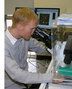
Photo: Biology professor Andrew Swan peers into the department's new confocal laser scanning microscope.
|
 New Microscope Provides Inside Look at Living Cells