![[Dr Reddish's Research Home page]](mcp-logo.gif) |
![[Dr Reddish's Research Home page]](mcp-logo.gif) |

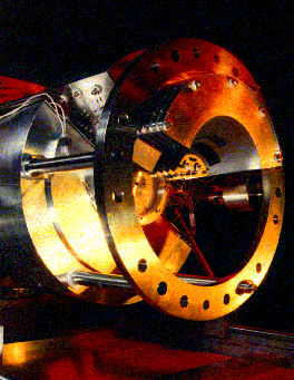
PDI is difficult to investigate experimentally because two outgoing particles need to be detected 'in coincidence' (i.e. time-correlated) to ensure the two electrons come from the same target atom/molecule. In this study the two ejected electrons are (independently) energy analysed during the detection process using electrostatic lenses which focus and steer the emitted electrons on to a 2-dimensional position-sensitive detector (see Fig.1). Novel toroidal analysers have been employed, which energy select the electrons while preserving the initial angle of emission. As a direct consequence of these focusing properties, the angular distribution of the photoelectrons are mapped directly onto the detectors and the resulting efficiency gains allow new experiments to be undertaken which otherwise would have been impossible. Experimental information is needed in all aspects of the PDI process; at Newcastle we have developed a spectrometer to investigate:
Figure 1: A schematic diagram showing the configuration of the
two toroids along with lines indicating central trajectories of electrons
with a selection of emission angles. The analysers are independent, i.e.:
they are able to detect dissimilar electron energies and resolutions. The
electron lenses are not shown for reasons of clarity. Electrons from the
interaction region are accelerated to the entrance of the toroid. The axial
symmetry results in the entrance lenses being slits on the curved surfaces
of a series of coaxial cylinders. Only electrons of a specific energy are
deflected through 142o to pass through the exit slit of the
toroid. The exit lenses, which consist of curved slits on the surfaces
of a series of coaxial cones, accelerate the electrons onto their respective
two-dimensional resistive anode encoders. The images on these detectors
are circular arcs with the circle centre on the photon axis.
For further details see: Reddish et al (1997) Rev. Sci. Instrum. 68 p2685