![[Dr Reddish's Research Home page]](mcp-logo.gif) |
![[Dr Reddish's Research Home page]](mcp-logo.gif) |
|
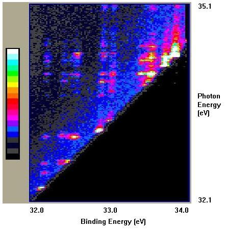
'Hot spots' Indicate Resonance Excitation. |
 |
![]()
Two-dimensional scanning in Atomic and Molecular Physics was first devised
for electron impact spectroscopy1, but has been more extensively
applied in photoelectron spectroscopy2,3. The basic concept
is to scan two variables (in this case - energies) over certain ranges
of interest and to view the variation in the cross section for the processes
covered by the 'area' scan. A large area covered by 2D scanning can only
be achieved when the spectrometer has a very efficient detection system,
such as provided by position-sensitive
detectors.
In the above photoionisation example, the binding energy range encloses the lowest-energy 'satellite' states of argon. [The large black 'triangular' region indicates that the photon energy is too low to excite states of that binding energy.] The blue verticle 'lines' show the variation in the cross section (with photon energy) of these excited ionic states. At certain photon energies the photon can also excite neutral Argon states in which two electrons are promoted. These 'autoionising' states subsequently decay by the emission of an electron, leaving the ions in a variety of excited states. These intermediate states are also called 'resonance' states as this additional mechanism for producing ionic states depends critically on the photon energy. The distribution of the ionic states to which the resonances decay depends on the details of the electron configurations of both the neutral and ionic states. In the states covered by the the figure, the most intense features are due to the resonant processes, showing their dominance of the cross section for producing excited, ionic states.
1 Reddish et al J Phys E: Sci. Instrum. (1988) 21 p203
2 For example see: Caffolla et al J Phys B: At. Mol. Opt. Phys. (1989) 22 L273
3 This method has been reviewed by Sokell et al J.
Elec. Spec. (1998) 94 107