 14
14
|
 14
14
|
| The spin of a nucleus can be compared to a gyroscope. 14 | |
MRIs make use of the unique property of atomic nuclei rotating in a strong magnetic field. These nuclei have a special "resonance" frequency that depends on the magnetic field. By absorbing radio waves of the same frequency, the nucleus' energy can be increased. Radio waves are re-emitted by the nuclei as they return to the lower energy state. The time it takes for the radio wave to do this is known as the ‘relaxation time’, and the different relaxation times result in varying bright and dark spots on the image.
Before the MRI scanning process can begin, patients must remove all metal objects, such as jewellery or watches, because they may interfere with the magnetic field. Once this has been done, the patient is instructed to lie on a bed and is placed into a magnetic field. The radio waves that are re-emitted by the nuclei in the patient’s body are detected by sensors which are placed around the body. Next, a computer uses ‘computed tomography’ techniques to assemble the slices into a three-dimensional image, allowing doctors to make a complete diagnosis.
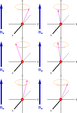
Larmor PrecessionNuclei have an intrinsic quantum property called spin. (Click here for a tutorial on quantum theory) When a magnetic field is imposed on the nucleus of an atom, this nuclear spin will orient itself according to this field, and so our z-axis can now be the direction of the magnetic field, for convenience.
The spin is represented by the arrow. Notice the tip of the arrow precesses similar to the top of the gyroscope. This spin allows absorption of a photon (a.k.a. light particle) of frequency νL, which is dependent on the strength of the magnetic field applied to the nulceus. This relationship is shown below:
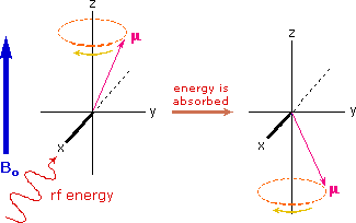
where γ (the gyromagnetic ratio) for hydrogen is 42.58 MHz/T. This special frequency is known as the ‘Larmor’ frequency (νL). If a really high magnetic field is present, this precession is in the radio-frequency (RF) portion of the spectrum. The atoms are placed in a non-uniform magnetic field. The nuclei of these atoms will have a different Larmor frequency of spin as a result of the equation above. As the radio frequency (RF) electromagnetic radiation (or light) is sent through the patient, the atomic nuclei in the body will absorb the energy. This absorption of energy causes nuclei to change the direction of their spin. You can intuitively understand this with the model shown above.
 14
14
|
|
The spin of nuclei flips when the nucleus absorbs a photon at its Larmor frequency. |
The RF signal has a frequency equal to the unique resonant frequency of the nuclei, the Larmor frequency. Once the RF signal is turned off, three processes occur, which are explained on the next page.
What properties of nuclei do MRIs chiefly use to create images?
A. Excitation Energy
B. Electron Spin
C. Nuclear Spin
What is the name of the unique frequency that nuclei spin at?
A. Nuclear frequency
B. Larmor frequency
C. Zeeman frequency