Technique | CAPSULE STAIN India Ink Method ,(/,)/,( |
Principle | The capsule or
glycocalyx is a gelatinous outer layer that is secreted
by the microbe and remains stuck to it. Capsules may be
polysaccharide, glycoproteins or polypeptides depedning
on the organism The large particles of the India ink are unable to penetration into the capsule |
Cautions | Any stray material in the
preparation that is not stained is frequently mistakenly
identified as a capsule 1: The film should be of the same
thickness as the capsulate organisms. |
Method | 1: Place a large loopful of
undiluted India ink on a slide 2: Mix into this a small portion of the bacterial colony or Mix a small loopful of the deposit from a centrifuged liquid culture. 3: Place a coverslip on top of the mixture. 4: Press down under a pad of blotting paper. |
Results | The capsule appears as a clear zone between the refractile cell outline and the dark background. |
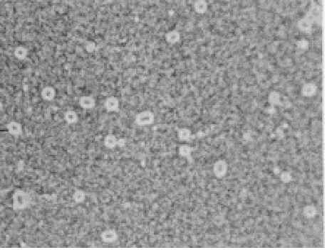
Positive control | Klebsiella pneumoniae (ATCC e13883) |
Negative control | Alacilgenes denitrificans (ATCC 15173) |
Reagents | Undiluted India ink Not all India Inks are suitable. Add 0.3% tricresol as a preservative |
Reference | J.P. Duguid 1951 J. Path Bact 63: 673 |
Technique | CAPSULE STAIN Antony's Method ,(/,)/,( |
Principle | The capsule or
glycocalyx is a gelatinous outer layer that is secreted
by the microbe and remains stuck to it. Capsules may be
polysaccharide, glycoproteins or polypeptides depedning
on the organism Both the cell and the capsule become stained by the crystal violet. However the crystal violet does not remain bound to the nonionic capsule and is easily dislodged with the copper sulfate solution, which also acts as a counterstain |
Cautions | DO NOT HEAT FIX
because there will be shrinkage Vigorous washing may dislodge the cells. |
Method | 1: Add a loopful of a
culture grown in either skimmed milk or litmus milk to a
microscope slide 2: spread the culture over the slide using the edge of a second microscope slide 3: Air dry 4: stain with 1% crystal violet for 2 minute 5: Wash with a solution of 20% copper sulfate 6: Blot dry 7: Examine under oil immersion |
Results | The cells are stained a deep blue or purple. The capsules are stained a light blue. The background make appear colourless or light violt depending on the degree of destaining |
Positive control | Klebsiella pneumoniae (ATCC e13883) |
Negative control | Alacilgenes denitrificans (ATCC 15173) |
Reagents | Crystal Violet 1gm/ 100ml distilled
water Copper Sulfate (CuSO4.5H20) 20gm/100 ml of distilled water |
Reference | Anthony,E.E. 1931 A note on capsule staining. Science 73:319 |
Technique | CAPSULE STAIN Hiss's Method ,(/,)/,( |
Principle | The capsule is a
gelatinous outer layer that is secreted by the microbe
and reamins stuck to it. Capsules maybe polysaccharide,
glycoproteins or polypeptides depending on the organism Both the cell and the capsule become stained by the crystal violet. However the crystal violet does not remain bound to the nonionic capsule and is easily dislodged with the copper sulfate solution, which also acts as a counterstain |
Cautions | Vigorous washing may
dislodge the cells. This method uses a dilute 0.1% crystal violet compared to the 1% crystal violet of Anthony's method |
Method | 1: Add a loopful of a
culture grown in either skimmed milk or litmus milk to a
microscope slide 2: spread the culture over the slide using the edge of a second microscope slide 3: Air dry 4: Heat FIX 5: stain with 0.1% crystal violet 6: Gently heat until steaiming (about 1 minute) 7: Wash with a solution of 20% copper sulfate 8: Blot Dry 9: Examine under oil immersion |
Results | The cells are stained a deep violet. The capsules are unstained against a purple background |
Positive control | Klebsiella pneumoniae (ATCC e13883) |
Negative control | Alacilgenes denitrificans (ATCC 15173) |
Reagents | Crystal Violet 0.1gm/ 100ml distilled water |
Reference | Hiss, P.J. Jr 1905 A contribution the the physiological differentiation of Pneumococcus and Streptococcus and to methods of staining capsules. J. Exptl Med 6: 317-345 |